
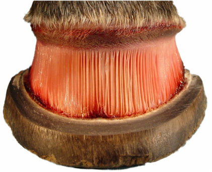
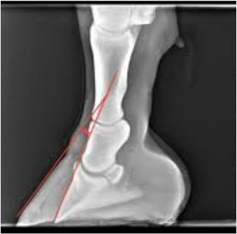
Laminitis is one of the most painful and serious hoof diseases in the equine industry. While the direct causes are not well known, there are multiple suggested predisposing factors that can trigger this disease. Laminitis or “founder” can best be described as problem where the “complex lamellar apparatus undergoes injury commonly leading to separation of the Lamellae dermales and epidermales” (Grundmann et al., 2014). Once the laminae layers become separated the distal phalanx (coffin bone) is essentially free in the hoof capsule. Because of the tension of the deep digital flexor tendon, which connects to the caudal aspect of the coffin bone, the entire bone can rotate downward, causing lameness and extreme pain. If the separation is throughout most of the hoof capsule, the coffin bone can sink, and this is nearly impossible to treat. This rotation can be detected in a radiograph of the affected foot. Once rotation of the coffin bone has occurred, there are no known ways to reverse the rotation, only stop it and prevent continual discomfort.
Causes of this condition can include an increased intake in highly soluble carbohydrates, often occurring after consuming from a very lush pasture or consuming too much grain. Once the soluble carbohydrates reach the hindgut, the increased fermentation leads to the death of the normal gram negative bacteria in the hindgut. Endotoxins are released from the cell walls of the bacteria. Once the endotoxins circulate through the body, due to the increased weight in the front legs, the endotoxins collect in the blood supply of the front hooves and damage the laminae, causing the separation of the coffin bone from the inside of the hoof capsule. This mechanism is only partially understood, but it is suggested that impaired blood supply to the hooves can also cause this separation. In a study of 22 ponies all subjected to increased sweet feed uptake it was discovered that “laminar COX-2 mRNA concentrations were increased […] Laminar COX-2 expression was immunohistochemically localised to multiple cell types (including epidermal keratinocytes, endothelial and fibroblast-like cells) […] This finding may implicate several cell types in the pathophysiology of endocrinopathic laminitis (Burns et al., 2014). Even with this finding the “signaling [that] likely to lead[s] to structural failure of the laminae in these animals appears to be distinct from the fulminant inflammation” (Burns et al., 2014), with an actual distinct cause still not being completely understood. In another study by Pass, S. Pollitt, and C.C. Pollitt glucose was found to be required for adhesion of basal epidermal cells to the basement membrane (1998). This adhesion was “maintained for more than one week when hoof explants were cultured in medium containing glucose, amino acids, vitamins, buffers and a variety of ions but for less than 2 days when cultured in physiological saline. [… But] when it [glucose] was added to saline, the explants remained intact for at least 2 days of incubation and in most instances up to 8 days” (Pass, S. Pollitt, and C.C. Pollitt, 1998). With a complete lack of glucose the separation of the tissues occurred, however the study did clarify that this is not evidence that laminitis will result (Pass, S. Pollitt, and C.C. Pollitt, 1998).
While most causes of this disease are related to nutrition, “road founder” can be caused by increased concussion on the hooves. After parturition, if the placenta is retained a mare may also develop laminitis.
While most causes of this disease are related to nutrition, “road founder” can be caused by increased concussion on the hooves. After parturition, if the placenta is retained a mare may also develop laminitis.
It is easy to recognize predisposing factors that can cause laminitis, some of which include ponies (due to their easy-keeping nature), obesity, insulin resistance, or the combination of the latter two, Equine Metabolic Syndrome. Horses that can be classified with any of the above should be monitored very closely in their diets, with nonstructural carbohydrates being avoided.
Treatment for this disease “varies with the severity of clinical signs and response to treatment; however, it tends to be an ad hoc approach” (Orsini, 2014). Phenylbutazone or “Bute” is “arguably the most widely used drug for the treatment of laminitis (Orsini, 2014). Due to the pain of this condition, it is most important to keep the horse comfortable, explaining the use of Bute, or other anti-inflammatory drugs. The frog must be supported either through bandaging the foot with a pad or having the horse stand in deep bedding or sand. When the coffin bone has rotated, creating a chronic case, the main goal of treatments would be to change the break-over point of the hoof and relieve pressure across the frog and sole. The hoof is reconstructed to elevate the heel and realign the hoof wall to the dorsal aspect of the coffin bone, making the two surfaces parallel once more.
Treatment for this disease “varies with the severity of clinical signs and response to treatment; however, it tends to be an ad hoc approach” (Orsini, 2014). Phenylbutazone or “Bute” is “arguably the most widely used drug for the treatment of laminitis (Orsini, 2014). Due to the pain of this condition, it is most important to keep the horse comfortable, explaining the use of Bute, or other anti-inflammatory drugs. The frog must be supported either through bandaging the foot with a pad or having the horse stand in deep bedding or sand. When the coffin bone has rotated, creating a chronic case, the main goal of treatments would be to change the break-over point of the hoof and relieve pressure across the frog and sole. The hoof is reconstructed to elevate the heel and realign the hoof wall to the dorsal aspect of the coffin bone, making the two surfaces parallel once more.

It is possible to prevent laminitis, when it is known the horse ingested an overload of soluble carbohydrates, by using cryotherapy, meaning the horse is stood in ice water for 48 hours, according to the study by Van Eps and Pollitt. The discomfort and weight avoidance “observed in the untreated limbs, was absent in the treated limbs” (Van Eps and Pollitt, 2004). It is hypothesized that the ice water caused the “vasoconstriction [that] may have prevented the delivery of these haematogenous [cytokines and hindgut bacterial products…] to the treated digit in this study” (Van Eps and Pollitt, 2004). On a day to day basis, it is best to feed susceptible horses diets low in nonstructural carbohydrates, or owners can soak the hay to decrease the amount of sugar. Pastures should also be limited or even avoided for susceptible horses. Obesity should also be avoided, so daily exercise or turnout can help keep horses healthy and could prevent this extremely painful disease.
*A majority of this section may be credited to Dr. Hess’ Laminitis Lecture.
Hess, Tanja, Dr. "Laminitis." ANEQ 346 Disease Management. Colorado State University, Fort Collins. 15 Oct. 2014. Lecture.
*A majority of this section may be credited to Dr. Hess’ Laminitis Lecture.
Hess, Tanja, Dr. "Laminitis." ANEQ 346 Disease Management. Colorado State University, Fort Collins. 15 Oct. 2014. Lecture.
