Have you ever heard this saying?

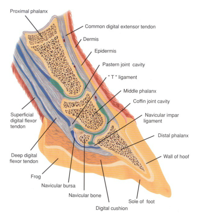
Any horse owner knows that the horse’s hooves are not just bones that are comparable to a human’s fingernails. The horse’s hoof is a capsule, containing the coffin bone (distal phalynx), navicular bone (distal sesamoid), digital cushion and the end of the deep digital flexor tendon. The hoof wall is composed of three layers: “the stratum externum, stratum medium, and stratum internum. The stratum medium is the thickest of the three layers and is characterized by its tubular and intertubular horn structure” (Pollitt, 2004). These three layers must support the horse’s body weight no matter what task he is doing, ranging from grazing in a pasture to running at 40 miles per hour on a track.
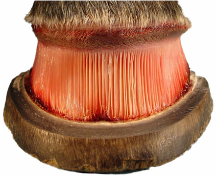
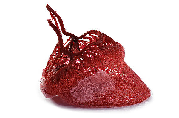
The hoof wall is composed of Keratin, with two groups found in the hoof wall, including “the ‘soft’ keratins of skin and the ‘hard’ keratins of horn” or hoof (Pollitt, 2004). The frog in the hoof is considered a member of the soft Keratin group, while the layers of the hoof wall are considered hard (Pollitt, 2004). The hoof is a structure that is continually growing from the keratinocytes located in the coronet band of the hoof, with an average growth of 1/4 to 3/8 inches per month (Wood). The equine hoof is a highly vascular structure, with a corium or dermis lying under the hoof wall, consisting of “arteries, veins and capillaries, and sensory and vasomotor nerves” (Pollitt, 2004). There is a corium specifically unique to the coronet band and one unique to the sole of the hoof.
Joining the hoof wall to the coffin bone is a complicated network called the “stratum lamellatum (layer of leaves) named after the 550 to 600 epidermal lamellae (primary epidermal lamellae) which project from its [the coffin bone’s] surface in parallel rows” (Pollitt, 2004). Lamellae (also called laminae) are rectangular in shape and cover the entire surface of the inner hoof wall.
These structures form a functional and healthy hoof. The links included on this webpage explain very common problems that impact racehorses. You will find an explanation of the problem, its likely causes, treatments and preventative measures.
|
|
